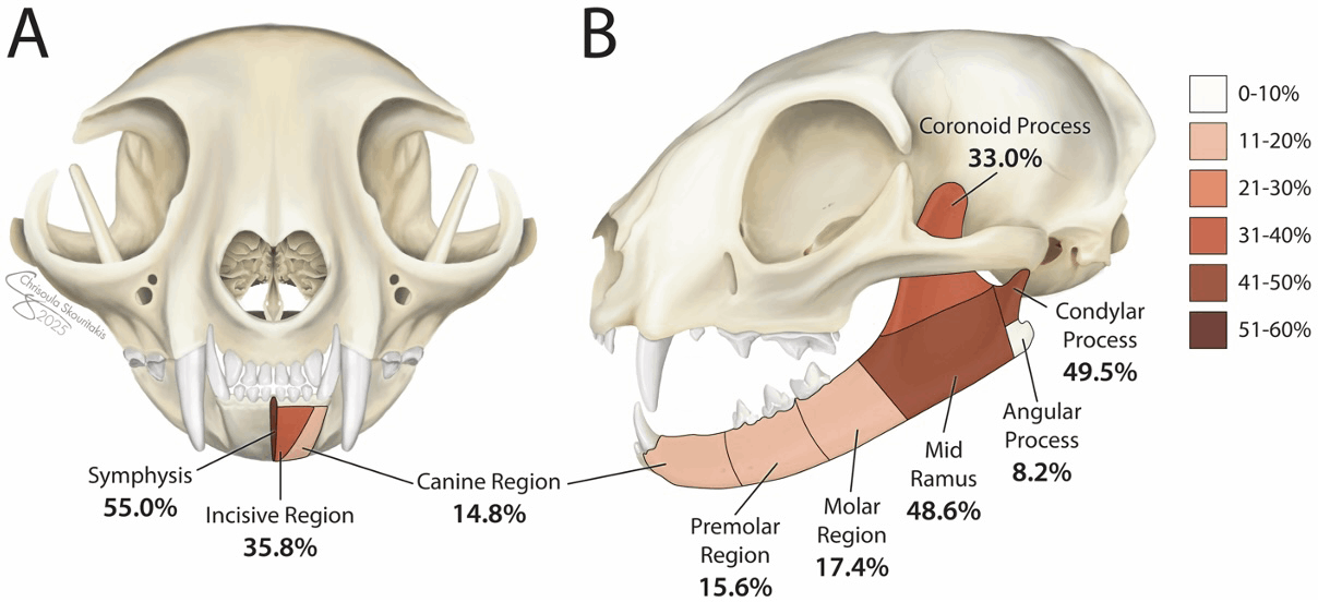
A retrospective study was performed on a population of 109 cats that were presented for evaluation and treatment of mandibular trauma. Medical records and diagnostic imaging were reviewed to determine mandibular fracture location, morphology, and treatment. Follow-up data were obtained from repeat clinical examination and diagnostic imaging. The most commonly injured anatomical locations were the mandibular symphysis (55.0%), the condylar process of the mandible (49.5%) and mid ramus (48.6%). More severe pre-operative fracture displacement was associated with a poor healing outcome in the mid ramus and coronoid process regions. The group of cats treated with open reduction and internal fixation (ORIF) had a significantly higher percentage of cats showing adequate healing (P = 0.0247) compared to the group of cats treated with maxillomandibular fixation (MMF). Cats treated with ORIF also had lower prevalence of persistent malocclusion (9.1%) when compared to cats treated with MMF (53.9%) (P = 0.0138, respectively). Placement of an esophageal feeding tube did not have a statistically significant impact on weight change in patients post-operatively (P = 0.0973). Patient survival was high at 94.5% indicating that cats that are treated for mandibular injuries have a good to excellent prognosis.