
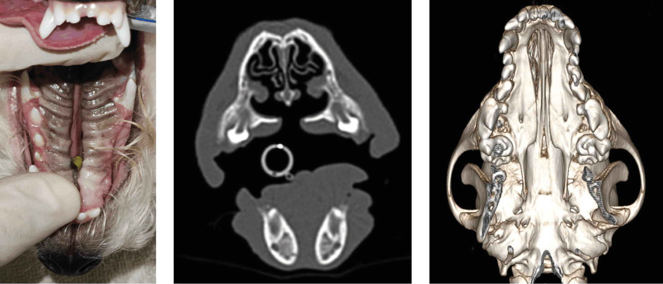
Cleft palate is one of the most common developmental malformations in dogs. It is an abnormal communication between the oral and nasal cavities due to the failure of fusion of the processes, bones and soft tissues responsible for forming the palate (Peralta et al.. 2018). Early diagnosis is essential and usually straightforward during a physical examination following birth (Evans and de Lahunta, 2020). Congenital oral and facial defects may manifest due to genetic and non-genetic factors in any dog breed, with a higher prevalence in some pure breeds and brachycephalic dogs. As facial and palatal development is complex and timely, this requires an orchestrated process. Depending on the timing of influencing factors, a primary and/or a secondary cleft palate may occur, which can manifest on its own or in combination (cleft lip, cleft palate, cleft lip and palate (Carlton and McGavin, 2016). The primary palate develops from the fusion of the medial nasal processes and the maxillary processes during embryonic development, forming the upper lip, alveolar crest and incisive bone (Kelly & Bardach, 2012). The secondary palate is formed by the midline fusion of the palatine processes originating from the maxillary processes. Fusion with the nasal septum (dorsally), palatine bones (caudally), and primary palate (rostrally) ensures complete separation of the oral and nasal cavities. Intramembranous ossification occurs in all but the caudal part of the secondary palate, which becomes the soft palate (Carlton and McGavin, 2016).
Primary cleft palates usually have limited clinical consequences, and surgical repair is often elective in the absence of clinical signs. In contrast, secondary clefts due to the large oro-nasal communication predispose affected puppies to malnutrition, aspiration pneumonia, and early mortality without supportive care and eventual surgery, typically after adult teeth eruption and before eight months of age. Understanding the embryological process of palate formation provides important context for the clinical differences between cleft types. It helps decision-making and setting client expectations regarding potential outcomes, as well as guiding treatment.
References
• Carlton, W. W., & McGavin, M. D. (2016). The alimentary system. In M. G. Maxie (Ed.), Jubb, Kennedy & Palmer’s pathology of domestic animals (6th ed., Vol. 2, pp. 1–157). Elsevier.
• Kelly, K. M., & Bardach, J. (2012). Biologic basis of cleft palate and palatal surgery. In F. J. M. Verstraete & M. J. Lommer (Eds.), Oral and maxillofacial surgery in dogs and cats (pp. 343–361). Saunders Elsevier.
• Evans, H. E., & de Lahunta, A. (2020). Miller’s Anatomy of the Dog (5th ed.). Elsevier.
• Peralta, S., Campbell, R. D., Fiani, N., Kan-Rohrer, K. H., & Verstraete, F. J. M. (2018). Outcomes of surgical repair of congenital palatal defects in dogs. Journal of the American Veterinary Medical Association, 253(11), 1445–1451. https://doi.org/10.2460/javma.253.11.144
